Home

canelură A trezi funcţie cilia electron microscopy ieri Temporar Simula

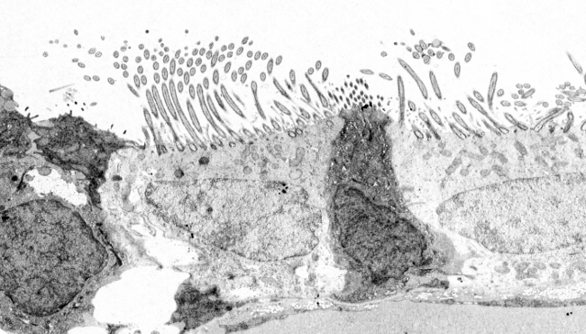
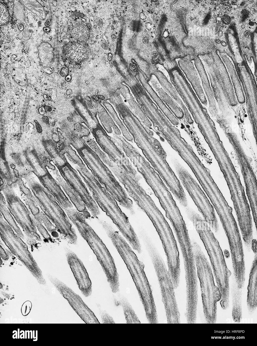
FocalPlane - It's #EMmonday! This is a transmission electron microscope  (TEM) image of murine tracheal ciliated cells. These cells contain a large  number of cilia that beat in a hydrodynamically synchronized manner
FocalPlane - It's #EMmonday! This is a transmission electron microscope (TEM) image of murine tracheal ciliated cells. These cells contain a large number of cilia that beat in a hydrodynamically synchronized manner

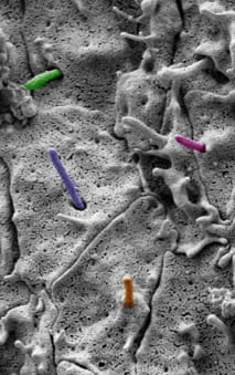
Visualizing renal primary cilia

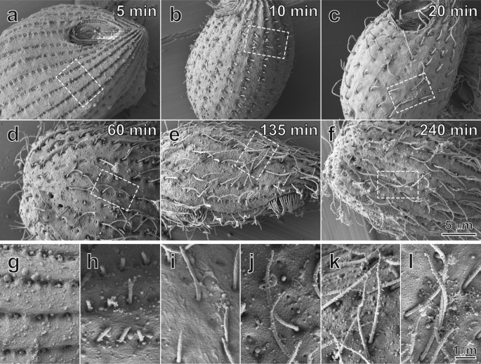
The Developmental Process of the Growing Motile Ciliary Tip Region |  Scientific Reports
The Developmental Process of the Growing Motile Ciliary Tip Region | Scientific Reports

Cilia, flagella, and centrioles | Celebrate Cytochemistry | Gwen V. Childs,  Ph.D.
Cilia, flagella, and centrioles | Celebrate Cytochemistry | Gwen V. Childs, Ph.D.

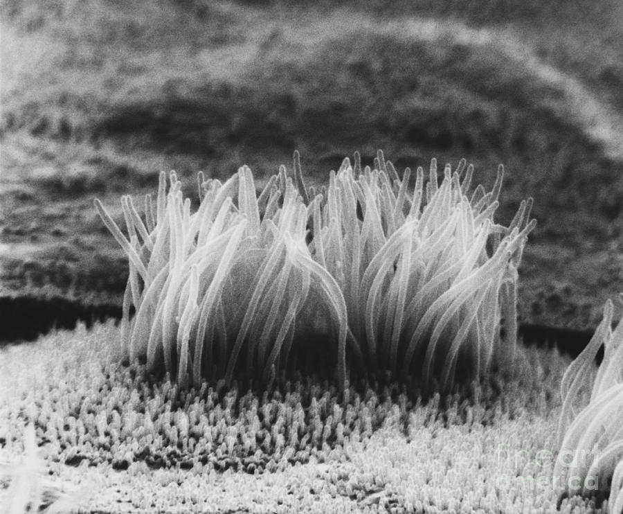
Mammalian Lung SEM | Microscopic photography, Things under a microscope,  Scanning electron microscope
Mammalian Lung SEM | Microscopic photography, Things under a microscope, Scanning electron microscope

The cell. More information. Cilia and flagella. Atlas of Plant and Animal  Histology
The cell. More information. Cilia and flagella. Atlas of Plant and Animal Histology

Electron Micrograph
Electron Micrograph

Electron microscopy of efferent ductule cilia. A.Higher magnification... |  Download Scientific Diagram
Electron microscopy of efferent ductule cilia. A.Higher magnification... | Download Scientific Diagram

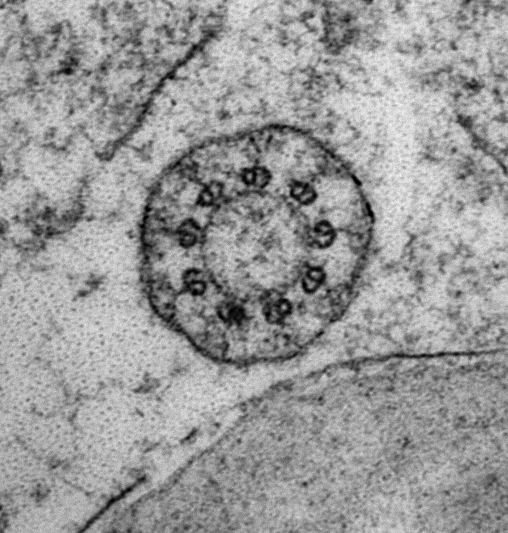
Electron microscopy findings showing normal cilia ultrastructure from... |  Download Scientific Diagram
Electron microscopy findings showing normal cilia ultrastructure from... | Download Scientific Diagram

A scientist wants to study the internal structure of cilia. Which electron  microscope would he use? - Quora
A scientist wants to study the internal structure of cilia. Which electron microscope would he use? - Quora
Electron Microscopy
Electron Microscopy

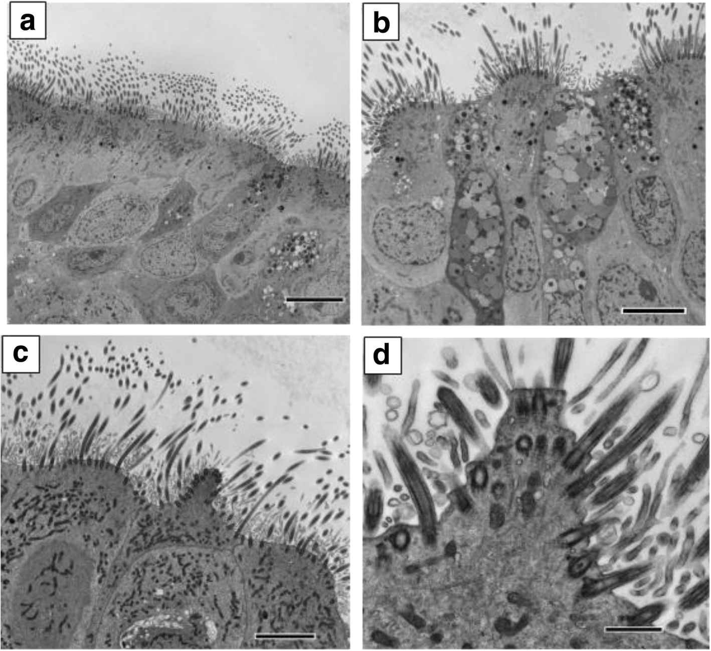
Ciliated conical epithelial cell protrusions point towards a diagnosis of  primary ciliary dyskinesia | Respiratory Research | Full Text
Ciliated conical epithelial cell protrusions point towards a diagnosis of primary ciliary dyskinesia | Respiratory Research | Full Text

Data | Free Full-Text | Transmission Electron Microscopy Tilt-Series Data  from In-Situ Chondrocyte Primary Cilia
Data | Free Full-Text | Transmission Electron Microscopy Tilt-Series Data from In-Situ Chondrocyte Primary Cilia
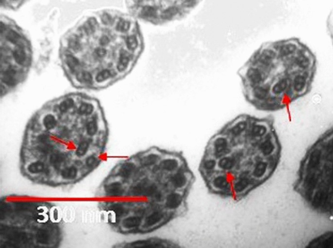
Transmission electron microscopy study of suspected primary ciliary  dyskinesia patients | Scientific Reports
Transmission electron microscopy study of suspected primary ciliary dyskinesia patients | Scientific Reports

Electron Microscopy
Electron Microscopy

Cilia Interpreting and Processing Molecular Signals | Brugmann Lab

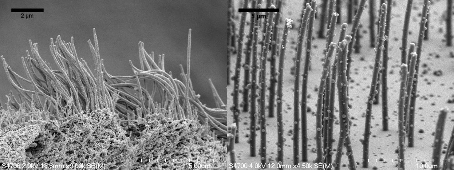
Biomimetic Cilia Arrays | CISMM

Characterization of Primary Cilia in Normal Fallopian Tube Epithelium and  Serous Tubal Intraepithelial Carcinoma
Characterization of Primary Cilia in Normal Fallopian Tube Epithelium and Serous Tubal Intraepithelial Carcinoma

Electron Microscopy of Flagella, Primary Cilia, and Intraflagellar  Transport in Flat-Embedded Cells - ScienceDirect
Electron Microscopy of Flagella, Primary Cilia, and Intraflagellar Transport in Flat-Embedded Cells - ScienceDirect

A 20-year experience of electron microscopy in the diagnosis of primary  ciliary dyskinesia | European Respiratory Society
A 20-year experience of electron microscopy in the diagnosis of primary ciliary dyskinesia | European Respiratory Society

RT TEM
RT TEM

Biomimetic cilia arrays generate simultaneous pumping and mixing regimes |  PNAS
Biomimetic cilia arrays generate simultaneous pumping and mixing regimes | PNAS

Cilia electron micrograph hi-res stock photography and images - Alamy

File:Cilia electron lobostoma.jpg - Wikimedia Commons

Sem Of Cilia On Epithelial Cells Photograph by David M. Phillips - Pixels

Images of cilia. ( a ) Scanning electron microscopy (SEM) image of... |  Download Scientific Diagram
Images of cilia. ( a ) Scanning electron microscopy (SEM) image of... | Download Scientific Diagram